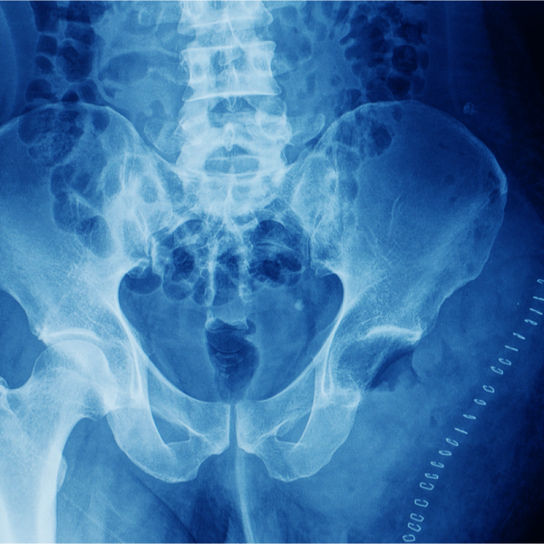
What is a chondrosarcoma?
A chondrosarcoma is a malignant bony tumour that arises from embryonic or mature cartilage tissue and develops mainly in the pelvis, hip or shoulder.
In only two percent of all cases, chondrosarcoma can also occur in the cranial region and manifests itself as headaches, double vision, hearing loss or tinnitus.
A chondrosarcoma usually grows rather slowly, but during its growth it destroys the bone and tends to form metastases quickly.
At just under 20 per cent, chondrosarcoma is the second most common type of bone cancer. People between the ages of 40 and 70 in particular have an increased risk of developing chondrosarcoma, with men being affected by the disease slightly more often than women.
What are the causes of chondrosarcoma?
The causes that lead to the development of chondrosarcoma are not yet sufficiently clear. In some cases, chondrosarcoma can occur as a result of previous radiotherapy used to treat cancer. However, people with certain hereditary diseases such as multiple cartilaginous exostosis and/or chondromatosis also have an increased risk of developing chondrosarcoma.
What forms is chondrosarcoma divided into?
The World Health Organisation (WHO) differentiates chondrosarcomas into the WHO categories of first grade (low), up to third grade (high), depending on their malignancy (malignancy). It is important to determine the severity of a chondrosarcoma, as this determines the subsequent form of therapy.
In addition, the following histological types of chondrosarcoma are distinguished, which can be classified according to the appearance of the cells and the localisation of the tumour:
- Conventional chondrosarcoma: occurs most frequently
- Clear cell chondrosarcoma: consists of cells that are visible under the microscope
- Mesenchymal chondrosarcoma: consists mainly of small tumour cells which have a high ratio of nucleus to cytoplasm
- Dedifferentiated chondrosarcoma: is a conventional chondrosarcoma that has transformed into a more aggressive type of cancer
What are the symptoms of chondrosarcoma?
Because chondrosarcoma usually grows slowly, it does not cause symptoms until the late stages of cancer. The symptoms of chondrosarcoma depend on where the tumour is located. In this case, the person mainly feels pain, especially when he or she does not move (pain at rest). In addition, swelling can develop in the area of the chondrosarcoma and the mobility of the affected body region can be limited. In general, chondrosarcoma can cause the following rather non-specific symptoms:
- Headache,
- Double vision,
- Hearing loss,
- Tinnitus,
- Cranial nerve deficits
How is chondrosarcoma diagnosed?
Chondrosarcoma, like other cancers, can be visualised using imaging techniques. In addition to magnetic resonance imaging (MRI), computer tomography (CT) can also be used for this purpose after a chondrosarcoma is suspected. In many cases, however, chondrosarcoma is diagnosed by chance. Within a CT, the chondrosarcoma is visible as erosion of the bones and often has calcium deposits. Within the MRI, the tumour has conspicuous honeycomb-like patterns.
However, an imaging procedure is not sufficient for a final diagnosis. The diagnosis of chondrosarcoma can only be made by a neuropathologist after a tissue sample has been taken and a fine-tissue examination has been carried out.
How is chondrosarcoma treated?
In most cases, an attempt is made to remove the chondrosarcoma by means of microsurgery. The latest technical procedures are used for this, such as so-called intraoperative neuromonitoring and neuronavigation. These procedures are designed to enable the neurosurgeon to remove the tumour as safely and precisely as possible.
The operation is often followed by radiotherapy, which is intended to improve the patient's survival rates. Doctors distinguish between the following options:
- Photon therapy: a beam of protons is used to specifically treat the tumour
- stereotactic radiosurgery: the treatment beams hit the tumour from different angles, thus affecting the healthy tissue as little as possible
- Proton therapy: beams of hydrogen atomic nuclei are used to treat the tumour in a targeted manner.
A study in the "European Journal of Cancer" has also shown that intensive chemotherapy, which is carried out in addition to surgery, can improve the chances of survival.
What is the aftercare for chondrosarcoma?
The treatment may be followed by a rehabilitation programme to help the patient return to his or her social and professional life. Within the rehabilitation programme, the person concerned should learn how to deal with the consequences of their cancer treatment. This is particularly necessary if the disease has led to an amputation, the wearing of a prosthesis or a nerve disorder, for example. Special sports programmes, which are offered in rehabilitation, are intended to help the patient to become physically healthy again. In addition, any psychosocial consequences can also be dealt with in therapy.
What is the prognosis for chondrosarcoma?
As with other cancers, the life expectancy of a chondrosarcoma depends on many factors. In addition to the tumour grade, its size and whether metastases have already formed, it also depends on the patient's general state of health. Patients who have had the tumour completely removed by surgery have a better chance of survival than patients who have residual tumour in their bodies. The 5-year survival rate after diagnosis is higher in patients who were treated with intensive radiotherapy after surgery than in patients who did not receive chemotherapy after surgery.
