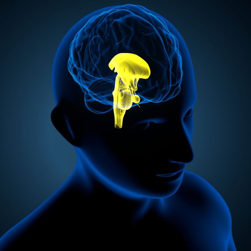
What is a craniopharyngeoma?
A craniopharyngeoma is a tumour that usually develops in the area of the stalk of the pituitary gland. Although the tumour is benign and slow growing, it can grow into the surrounding structure such as the pituitary gland (gland at the base of the brain), the pituitary stalk (glandular body of the pituitary gland and the hypothalamus), the optic nerves and/or the vessels. Between 2.5 and 4 percent of all brain tumours are craniopharyngeomas, about half of which occur in childhood. An above-average number of craniopharyngeomas develop between the ages of 5 and 10. Adults often develop a craniopharyngeoma from the age of 40, although men and women can be affected in roughly the same way .
What causes a craniopharyngeoma to develop?
A craniopharyngeoma forms through the sudden proliferation of residual cells of the Rathke's pouch. The Rathke's pouch describes a structure that originates from the embryonic development of the pituitary gland and usually regresses. In most cases, craniopharyngeomas grow in the area of the pituitary stalk. From here, the tumour can spread in different directions. About 5 percent of all craniopharyngeomas form purely intraventricularly (within the ventricle). In craniopharyngeoma, the third ventricle is particularly often affected.
What are the different types of craniopharyngeoma?
The following two types of craniopharyngeoma are commonly distinguished:
- adamantinous Craniopharyngeoma: which is often calcified and hard and has a higher irritidv risk. It occurs more often in children.
- papillary Craniopharyngeoma: which is extremely rarely calcified and has a lower risk of recurrence. It occurs mainly in adults.
What symptoms can be caused by a craniopharyngeoma?
Since the craniopharyngeoma forms in close proximity to the optic nerves, the hyophysis, the pituitary stalk, the hypothalamus as well as the brain stem, among others, the tumour can lead to the following symptoms:
- Headaches,
- Headaches, visual field disturbances and/or visual disturbances,
- hormonal disturbances, which can especially trigger diabetes insipidus, which manifests itself through increased thirst,
- Nausea and/or vomiting,
- neurocognitive disorders,
- hypothalamic disorders, which is mainly manifested by weight gain,
- in children, absence of puberty or other growth disorders.
As a rule, craniopharyngeoma does not cause any complaints in adults, especially in the initial phase
. If headaches occur, but also
visual disturbances, these can indicate a larger growth of the
tumour in the pituitary region.
How is a craniopharyngeoma diagnosed?
A craniopharyngeoma can usually be diagnosed using the imaging technique of magnetic resonance imaging (MRI). In this procedure, the patient is given a contrast medium so that the MRI can show both the location of the tumour and the extent of the tumour. In individual cases, a computer tomography (CT) can also be carried out to assess whether tumour calcifications are present. It is also advisable to have an endocrinological examination carried out if a craniopharyngeoma is suspected. This can help to identify and treat possible hormonal disorders. If the MRI scan shows that the tumour is in contact with the optic chiasm (important section of the visual pathway), an ophthalmological check is also necessary. In the majority of all cases of the disease in adults, signs of a hormone deficiency as well as psychological changes can already be detected at the time of diagnosis.
How is a craniopharyngeoma treated?
Because craniopharyngiomas can expand as they grow, displacing adjacent structures and causing permanent brain damage , the first choice of treatment is always complete surgical removal of the tumour. Doctors can choose between the transcranial approach and the transnasal transsphenoidal approach . While the transcranial approach involves opening the skull to microsurgically remove the tumour, the transnasal transsphenoidal approach involves removing the tumour through the nose.
If a complete resection is not possible due to the location of the tumour , postoperative radiotherapy or radiosurgery can be performed to reduce the risk of the tumour developing again (recurrence).
Research is currently being done to develop chemotherapy as a complementary treatment option. In addition , drugs for the therapy of papillary craniopharyngeoma are being tested. These are so-called BRAF inhibitors, which include vemurafenib and dabrafenib.
What complications can occur?
After the surgical removal of the tumour, there can mainly be disturbances in the electrolyte and hormone balance. In the best case, these disturbances are only temporary. However, it can also happen that the disturbances remain permanent. Especially after the operation, the blood values and the water balance of the patient are therefore closely monitored in order to be able to compensate for the hormone deficiency with medication if necessary.
What is the aftercare for a craniopharyngeoma?
Due to the risk of recurrence of craniopharyngeomas, the patient requires lifelong follow-up care . This includes clinical and examinations, as well as a check-up on the patient's condition. In addition to clinical and endocrinological examinations, this also includes MRI checks, which should initially be carried out at annual intervals. In addition, the sella region in particular should be regularly checked by an ophthalmologist . If the tumour develops again, it should be surgically removed at an early stage and, if necessary, the remaining tumour cells should be irradiated afterwards.
In patients in whom the tumour has already grown into the neighbouring brain structures, there may be a loss of control of possibly vital functions. The quality of life of those affected is significantly reduced as a result. In 30 percent of all cases, there may also be a high degree of obesity , which can lead to diabetes and heart disease . As part of medical monitoring, the overweight should be reduced in order to improve the long-term quality of life of the patients.
