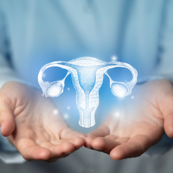
What is a leiomyoma?
Leiomyomas are among the most common benign tumours of the uterus and arise from the muscle cells of the myometrium. A leiomyoma can occur sporadically as a so-called myoma uteri or muliply as uterus myomatosus. Particularly small leiomyomas are called myoma germs. In 40 to 50 percent of all cases, leiomyomas occur in women who are older than 30 years. They are most often found in the uterine corpus (body of the uterus) and less often in the cervix (lowest point of the uterus). In a few cases, leiomyomas also form in the broad uterine ligament (ligamentum latum) or in the fallopian tubes. Because leiomyomas can grow to a considerable size, up to 10 cm in diameter or more, they can contribute to a deformation and enlargement of the uterus.
What are the different types of leiomyomas?
Leiomyomas are classified according to their location as follows:
- subserosal myoma: located below the serosa, can produce stalk-like fibroids which can lead to haemorrhagic infarction.
- intramural myoma: located in the uterine wall,
- submucosal myoma: grows below the lining of the uterus, often penetrates into the uterine cavity and can cause bleeding. In some cases, ascending infections can also be favoured by a submucosal leiomyoma.
Subserous and intramural fibroids are usually removed through an abdominal incision or laparoscopy. A submucosal myoma, on the other hand, is removed through the vagina by hysteroscopy.
What causes leiomyomas?
Leiomyomas are hormone-dependent tumours and usually grow slowly. They develop from smooth muscle cells that are found in the internal organs, for example in the uterus, kidneys or gastrointestinal tract.
Are leiomyomas dangerous during pregnancy?
Due to hormonal changes, leiomyomas can increase in size during pregnancy, which can lead to various complications. For example, having a leiomyoma during pregnancy can increase the risk of premature birth. This is because leiomyomas can cause the placenta to adhere incorrectly, leading to spontaneous abortion. But the risk of premature labour or positional abnormalities of the baby also increases. Studies show that long-term use of ovulation inhibitors can reduce the likelihood of developing a leiomyoma.
What symptoms does a leiomyoma cause?
In many cases, a leiomyoma does not cause any symptoms. The most common symptoms, if they occur, are bleeding disorders in the form of menstruation that is too heavy and lasts too long (menorrhagia), bleeding outside of the period (metrorrhagia) or increased menstrual bleeding with very heavy blood loss (hypermenorrhoea). The following symptoms tend to occur less frequently:
- strong urge to urinate,
- Pain during sexual intercourse,
- Lower abdominal pain,
- swollen abdomen, especially with large leioymomas,
- Constipation,
- Back and/or leg pain,
- Kidney or side pain
How is a leiomyoma diagnosed?
After taking the medical history, the gynaecologist will perform a palpation examination. This is usually done through the vagina, but also through the abdominal wall and rectum. If there is a larger leiomyoma or several leiomyomas, they can usually be felt. In addition to this palpation, a vaginal sonography (ultrasound examination) is performed. A myoma can be distinguished in the ultrasound image by its echo arms, rounded appearance and smooth borders. Sonography can be used to determine the exact location and size of the leiomyoma.
If the leiomyoma is pressing on the ureter, the kidneys and the urinary tract are usually also examined either by ultrasound or by X-ray with contrast medium (pyelogram). If the results of the examination are unclear, the gynaecologist can order a magnetic resonance imaging (MRT) and, if necessary, also have a blood test and a measurement of the hormone level. The latter examination is mainly done if there is a suspicion of anaemia.
How is a leiomyoma treated?
A leiomyoma can be removed surgically. The following therapeutic procedures are available, which differ from each other as follows:
- Myoma enucleation: The fibroids are removed from the uterus without affecting it. The myoma enucleation procedure is used in particular in cases where there is a desire to have children. The surgical procedure is carried out by means of an abdominal incision, laparoscopy or hysteroscopy. Which procedure is used depends on the exact location and size of the leiomyoma. In some cases, the leiomyoma can form again after myoma enucleation.
- Myoma embolisation: cuts off the blood supply to the leiomyoma in order to shrink it in this way. Mymembolisation can be done as an alternative to surgery to remove either the leiomyoma (myomectomy) or the uterus (hysterectomy).
- Hysterectomy: describes the complete or partial removal of the uterus.
In addition, drug treatments can also be used for a leiomyoma. For example, GnRH analogues can be given for this purpose. These are drugs that artificially lower the oestrogen level in order to inhibit the growth of a leiomyoma. The insertion of a hormonal IUD also has the same purpose.
What is the course of the disease and the prognosis for a leiomyoma?
Only leiomyomas that cause symptoms need treatment. If left untreated, they can cause chronic lower abdominal pain. If the patient has not yet finished planning for a child, conservative forms of therapy should be given priority over removal of the uterus (hysterectomy). Patients without symptoms should attend regular check-ups. Even benign leiomyomas can cause complications such as urinary tract infections or functional disorders of the bladder, intestines or kidneys. Even after successful treatment, leiomyomas can form again.
Can a leiomyoma become malignant?
It is quite rare for benign fibroids to degenerate. This is only the case in less than one percent of all cases and is almost exclusively observed in women in or after menopause.
