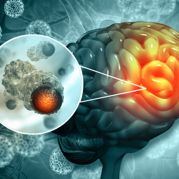
What are malignant meningiomas?
A malignant meningioma is a malignant brain tumour that occurs rather rarely and accounts for only 2 to 3 percent of all meningiomas. Malignant meningiomas form in the meninges of the brain, and men are more likely to develop this disease than women. Malignant brain tumours grow rapidly in size and can grow into the surrounding brain structures or into another tissue through the spread of daughter tumours (metastases). The prognosis for a malignant meningioma is rather unfavourable and always requires postoperative radiotherapy.
What is the severity of a malignant meningioma?
The World Health Organisation (WHO) distinguishes between three tumour grades, whereby the first two grades describe a benign brain tumour. A malignant meningioma manifests itself in the third and most serious tumour grade:
- WHO grade III: anaplastic men ingioma is the rarest form of all meningiomas and accounts for only about 2 per cent of all cases. An analplastic meningioma can form metastases and thus spread to other organ structures.
Meningiomas of the second WHO grade are also not only difficult to treat in some cases, but can even become malignant. Surgery can be more difficult than for meningiomas of the first WHO grade.
How does a malignant meningioma develop?
As with other types of cancer, a malignant meningioma develops when a certain type of cell degenerates. Although the exact cause of this mutation is still unclear to medical experts, they assume, especially in the case of the development of a malignant meningioma, that children who have received radiation therapy due to cancer, but also atomic bomb survivors, have a greater risk of developing a malignant brain tumour (an anaplastic meningioma).
What are the symptoms of a malignant meningioma?
Due to its rapid growth, a malignant meningioma can cause seizures. However, particularly large tumours can also cause the following neurological disorders:
- Speech disorders,
- Paralysis,
- Visual disturbances,
- Deterioration of the sense of smell,
- in some cases, personality changes can even occur.
Depending on where the meningioma occurs, it can also affect the drainage of cerebrospinal fluid. This can lead to a condition called hydrocephalus. Headaches are also a common side effect of meningioma, but are rarely caused directly by the tumour.
How is a meningioma diagnosed?
Because a malignant meningioma usually already causes symptoms, the diagnosis at this stage is usually no longer an incidental finding. The patient presents to the doctor because of the symptoms, whereupon a magnetic resonance tomography (MRT) or a computer tomography (CT) is carried out. A contrast agent can be used to make abnormalities visible. Both an MRI and a CT scan can show the exact position and size of the tumour. An additional X-ray examination of the blood vessels of the head (angiography) can also determine which vessels are connected to the tumour or whether certain vessels have been displaced by the tumour and thus restrict the blood flow.
How is a malignant meningioma treated?
Malignant meningiomas usually grow continuously and compress the brain due to their size. For this reason, they usually cause symptoms and should be treated urgently. Surgical removal of the tumour is the standard treatment. However, since this is not always possible due to the localisation and size of the tumour, radiosurgical therapy may also be considered. The respective treatment therefore depends on the location of the tumour, but also its size and speed of growth. There are also many other factors, such as the patient's general state of health.
In an operation, the meningioma is reduced in size from the inside. In this way, the borders to the neighbouring tissue are relieved. Surgically, the tumour-bearing meninges are removed and replaced. The process of meninges replacement can vary in intensity from patient to patient. This depends on whether the tumour has a smooth border or has already infiltrated the brain surface. The intervention becomes particularly difficult if the tumour is already supplied with blood by normal brain vessels. Thanks to the latest technical procedures, such as augmented reality, so-called neuronavigation or intraoperative neuromonitoring, it is possible for the neurosurgeon to look into the tissue with maximum precision and to perform the intervention under the greatest safety precautions.
All meningiomas are sensitive to radiation. For a meningioma that cannot be operated on, radiation therapy is therefore usually performed. However, the tumour must not have exceeded a certain size for radiotherapy. If neither surgery nor radiotherapy is an option, so-called radionuclide therapy is an alternative. This is mainly used in difficult cases with progressive progression of the disease. In radionuclide therapy, the tumour is targeted with radioactive drugs. The so-called radiopharmaceutical is often used here. This is a radioactive substance that binds special receptors (somatostatin receptors) on the surface of the tumour, where it has a local radiation effect and destroys the tumour cells.
In general, it can be stated with regard to the treatment of a malignant meningioma that a complete removal of the tumour is almost impossible from the second WHO grade. Depending on where the meningioma is located, it can affect important structures. This is the case, for example, if the meningioma is located near the pituitary gland or the brain stem. This is where not only the aorta, which is responsible for supplying blood to the brain, is located, but also the and the pituitary gland.
What is the prognosis for a malignant meningioma?
The chances of a complete cure for a malignant meningioma of the third degree of severity are rather unfavourable. This is mainly because the brain tumour can metastasise.
