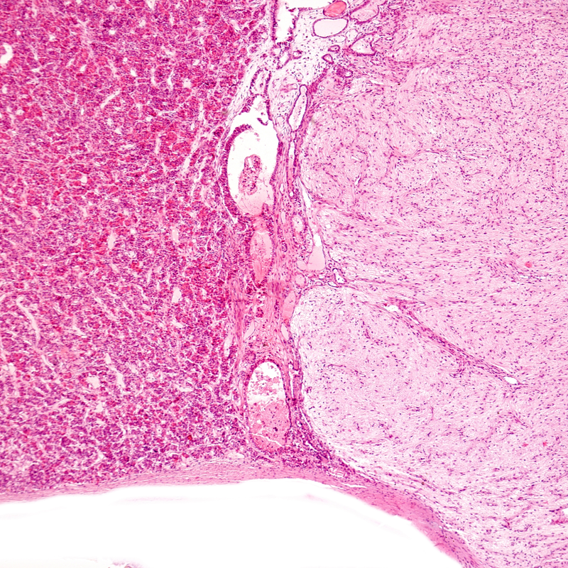
What is a Rathke's pouch tumour?
A Rathke Pouch Tumour is a benign space-occupying tumour that originates from the remnants of the Rathke pouch. The Rathke pouch tumour, also known in German as Rathke Taschen Tumor, owes its name to the German anatomist Martin Heinrich Rathke. The following types are distinguished:
- The Rathke cyst: it is filled with fluid , surrounded by epithelium and usually has no clinical significance . However, depending on the size of the Rathke's cyst, symptoms of a pituitary tumour are possible.
- The colloid cyst: it is filled with a viscous mucous fluid - The craniopharyngeoma
How common is a Rathke pouch tumour?
Especially Rathke cysts are often only discovered by chance, although they are not rare at all . The prevalence is around 13-23 %, whereby women are affected more often than men. This results in a ratio of 2:1.
How does the Rathke pouch tumour develop?
The Rathke's pouch is a so-called protrusion of the roof of the throat. In the fetus, the anterior pituitary gland develops from this pouch. Thus this is not a brain tissue, but like the CNS it has an ectodermal origin. In further development, this protuberance is strangulated and thus loses its connection to the oral cavity. This resulting cavity of the bay forms the pituitary vesicle. Normally, it forms back again completely. However, in rare cases, derived cysts can be found between the Paris intermedia and Paris distalis of the pituitary gland. These are then called Rathke cysts. A benign tumour originating from the Rathke's pouch is the craniopharyngeoma.
What are the symptoms of a Rathke's pouch tumour?
The most common type of Rathke's pouch tumour is the Rathke's cyst. Due to their usually very small size, these do not cause any symptoms . However, if they grow, they can put pressure on the adjacent areas of the brain. This can cause the following symptoms :
- Visual disturbances and/or also restrictions of the visual field due to compression of the optic nerves
- Hormonal imbalances due to compression of the pituitary gland
- In children, symptoms such as pubertas praecox, growth retardation and diabetes insipidus may occur.
How is a Rathke's pouch tumour diagnosed?
On the basis of an MRI image with contrast medium shows predominantly a small Rathke's cyst, which is located in the area of the pituitary gland and usually has no suprasellar extension. MRI or MRI is the first choice in the diagnosis of Rathke's pouch tumour. The MRI image can provide important information about the extent of the cyst and the location. If patients have a larger cyst, an endocrinological evaluation should be carried out so that any disruptions of the hormone balance can be diagnosed and treated. If the MRI image shows a cyst in contact with the optic chiasm, an additional eye examination will be organised and performed. The ophthalmologist can detect and quantify visual field and visual impairment . These often go unnoticed for a long time by those affected .
The same examination scheme is also given and recommended for colloid cysts and craniopharyngeoma.
How is a Rathke's pouch tumour treated?
In the case of small Rathke's cysts that do not cause any symptoms, it is recommended to check their progress with the help of an MRI at intervals of 6 to 12 months. This follow-up is important to be able to exclude an increase in size. In the case of tiny cysts that are smaller than 5 mm, no check is necessary. Unless symptoms develop.
However, if a Rathke's cyst causes symptoms and discomfort, surgical therapy is necessary. As a rule, the surgical approach is via a transnasal, transsphenoidal approach. The procedure can be performed microsurgically, fully endoscopically or endoscopically assisted. As the wall of the cyst is often attached to surrounding structures such as the pituitary stalk or optic chiasm, fenestration or partial resection of the cyst is recommended.
Also surgery is indicated when the Rathke's cyst exerts compression on the optic chiasm and there is visual field disturbance and/or visual disturbance. In children, the benefit must be weighed against the risk. If the headache is so severe that the child's quality of life suffers, surgery is usually indicated. Doctors use clinical judgement here. However, since the headache persists in one third of all patients operated on in childhood , the decision for an operation must be evaluated with caution in view of the symptoms alone . If one considers the results from studies in which it has been proven that approx. 31-35 % of the cysts even regressed in size, i.e. decreased in size , a conservative therapy in the sense of "wait and scan" is more likely to be recommended.
In the case of colloid cysts, the above-mentioned treatment is also recommended in this way. In the case of a craniophayngeoma , surgery is always the first choice of therapy.
What are the risks of surgery for a Rathke pouch tumour?
Possible complications after resection of Rathke's cysts are primarily considered to be disturbances of the hormone balance and the electrolyte balance. For the most part, however, these disturbances are only temporary. In view of these disturbances, close monitoring of blood values and water balance after the operation is essential. In some cases, it is necessary to substitute hormones with medication . A Rathke's cyst that recurs is not uncommon, therefore in most cases another operation will have to be repeated in the course of time.
What is the prognosis for a Rathke pouch tumour?
Since a Rathke's pouch tumour is usually benign, the prognosis is also very good. Nevertheless, the growth of a Rathke cyst, a colloid cyst or a craniopharyngeoma should be observed at regular intervals . Regular imaging by MRI or MRT is also recommended to exclude possible recurrences.
