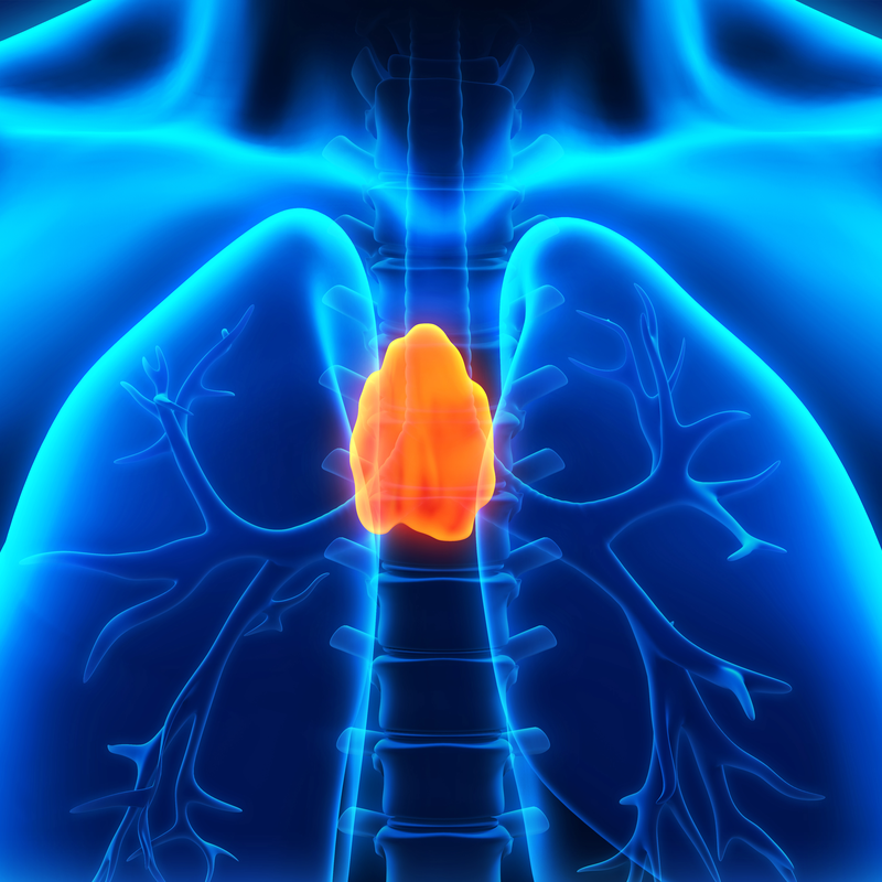
What is a thymoma?
Thymoma is a rarely occurring tumour of the thymus, or more precisely of the thymus gland. In three quarters of cases, thymomas are benign. One quarter, however, are malignant. They are divided into malignant thymomas and thymic tumours according to their appearance and the degree of differentiation of the diseased cells and the tendency to spread.
Like the benign thymomas, the malignant thymomas also develop on the surface of the thymus gland. However, in the benign thymomas, the thymic cells multiply much more slowly and also do not settle outside the organ. In malignant thymomas, the cells grow much faster and also invade the surrounding tissue. Preferably the cells of malignant thymomas spread in the lymphatic channels of the thoracic cavity. The cells of thymic tumours also settle in the organs further away and usually form metastases there.
How does a thymoma form?
The thymus forms the central organ of the lymphatic system. The tyhmus is located in the mediastinum in front of the heart and is most important for the selection of T lymphocytes. In about 50 % of cases, the thymoma is causative for the formation of a tumour in the anterior mediastinum.
How common is a thymoma?
Thymomas occur rather rarely. They only make up about 0.2 to 1.5 % of all tumours. In Germany, about 0.2 to 0.4 of every 100,000 people get a thymoma. Women and men are equally affected. Thymomas can occur at any age, although the most common occur between the ages of 50 and 60.
What are the symptoms of a thymoma?
In most cases, it is only when the thymoma is more advanced that it causes symptoms. . About 30 % of patients do not show any symptoms when they are diagnosed. As a rule, they only appear when the thymoma grows into the surrounding tissue or increasingly displaces surrounding structures. About 40 % of the symptoms are caused by the large tumour mass in the chest. The most common symptoms are cough, severe shortness of breath and chest pain.
Other possible symptoms:
- Difficulty swallowing
- Hoarseness due to paralysis of the cervical nerve -
- Heart dysfunction if the mass of the tumour presses on the heart.
How is a thymoma diagnosed?
In many cases, the diagnosis of thymoma tends to be an incidental finding, which is made during an X-ray examination of the chest. However, even if symptoms point to the suspicion of a thymoma, the doctor will first record the patient's current complaints, medical history and any risk factors. This will be followed by a thorough physical examination. This can give the doctor important information about the nature of the disease. If it has not already been done, this is followed by an X-ray examination of the thorax. A blood test and urine test are also carried out. On the basis of a CT and MRI examination , the size, location and extent of the tumour can be determined. In this way, the stage of the thymoma can also be determined.
- Stage I: Tumour within the thymus gland, in the thymus capsule
- Stage II: Spread of the tumour to surrounding fat or the lining of the lung cavity
- Stage III: Spread of the tumour to organs near the thymus
- Stage IVa: Spread of the tumour to the pleura of the heart or lung
- Stage IVb: Spread of the tumour through blood or lymph vessels
Stage I is considered a non-invasive malignant thymoma. Stages II-IV old as invasive thymic carcinoma. Of course, the stage of the tumour always depends on the type of therapy.
How is a thymoma treated?
The absolute gold standard for the treatment of thymomas is surgery. From the oncologists' point of view, the removal of the tumour is the most important factor for the survival of the patients. This means that all structures attached to the thymoma must also be removed, which includes both the residual thymus tissue, the surrounding lymph nodes as well as all fatty tissue components and connective tissue components. The chance of complete removal of the tumour decreases the further the stage has progressed. In patients with an encapsulated stage I tumour, it can be completely removed in 100 % of cases . In the case of a larger tumour, i.e. stages II-III and even after incomplete removal, an additional radiotherapy can be considered. In the case of stage IV thymomas, removal of the tumour is only possible in 30 % of cases, and metastases have already formed in stage IV.
An operation can be carried out via different access routes and depends on the size of the tumour and the extent of the tumour. In most cases, it is necessary to open the breastbone. If the view is good, all the affected tissue and the lymph nodes can be removed. In some cases, a lateral opening of the chest is also possible. If the thymoma is still tiny, the keyhole technique is also possible.
How is a thymoma followed up?
The patient usually stays in the hospital for about twelve days after an open operation. After eight weeks, the chest can be fully loaded again . Since all thymomas are considered malignant, follow-up care is essential. Thymomas have a very high local recurrence rate, because new tumours can still develop up to 10 years after an operation. Attention must also be paid to secondary tumours such as non-Hodgkin's lymphoma and soft tissue sarcomas. It is therefore important that the patient has a quarterly check-up at for the first twelve years after a successful operation . After that, a CT chest every twelve months is important. Ideally, follow-up can be done with the thoracic surgeon who first treated the patient.
What is the prognosis for thymoma?
The prognosis for thymomas is good overall, but best if the tumour could be completely removed. In some cases, this may not be possible , but the operation is still useful because the survival of the patient can be significantly improved by a reduction of the tumour mass . Afterwards, however, it is imperative that follow-up radiotherapy be carried out. The 5-year survival rate is quite high at approx. 80 % . Overall, there are many important factors that can have a significant positive influence on survival. One is the complete removal of the tumour in the good and the other is a low stage and the presence of the thymoma in the capsule.
